X-ray examination

 Dental x-ray, also known as radiogram is an auxiliary diagnostic and therapeutic tool. It assesses the dental crown and root condition, also the structure of the tooth: periodontal ligament, alveolus outgrowth – a bone.
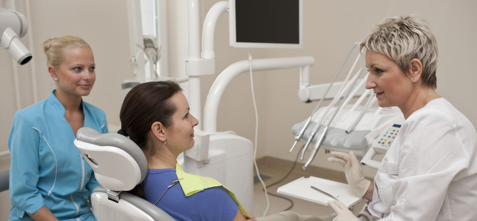
Today, dental x-rays are a very important supportive treatment tool. High-quality photos of teeth allow dentists to assess the exact condition and plan the patient’s treatment: root canal preparation and filling, dental implantation, prosthodontics, tooth extraction and other complex procedures. Sometimes without dental photos it is impossible to accurately assess the interdental or root caries, root tip inflammation, tooth bone disintegration.
Our clinic carries out dental x-rays, which allow very detailed assessment of patients’ condition. Currently the clinic has the ability to perform a dental digital radiogram which specific program helps to make an accurate diagnosis. Seeing such a photo, specialist can determine the length of the root canals and their formation, the length of periodontal pockets, tooth decay, etc.
Radiological examination is carried out by special certified x-ray device, which is checked for radiation safety and meets all the requirements. It emits a very small and narrow x-ray beam, which makes minimal exposure, and lead protection preserves the closest organs from irradiation. Therefore, this study is safe. Nowadays, digital technology makes it possible to reduce x-ray dose to tens of percent. Thus, it is a myth, that during radiographic studies, patients get a high dose of x- rays.